|
|
|
Checklist of Sea Anemones (Cnidaria: Anthozoa) From
Presented by Dr. Fayez F. A. Shoukr, Professor of
Invertebrates, Zoology Department, Faculty of Science,
Shoukr, F. A.,
Mona, M.H. and Badawy, B.E. (2000) : Taxonomy Of Some
Sea Anemones (Cnidaria: Anthozoa)
From
ABSTRACT:The
taxonomic description of the fouling anemone Aiptasia
diaphana (Cnidaria: Anthozoa) is firstly given in
INTRODUCTION
Sea
anemones are marine invertebrates which belong to Order Actiniaria,
Sub-Class Zoantharia, Class Anthozoa
and Phylum Cnidaria (Coelenterata).
They are mainly sessile with some swimming forms. They have unique cellular
weapons called nematocysts. The
anemones occupy a wide range of habitats among marine fouling communities,
coral reefs and rocky shores especially at the intertidal
zones.
Indeed, the
early studies on anemone taxonomy of
The local anemones have attracted the attention of
many workers in Egypt for their beautiful colours, ecological relationships
with other marine organisms (fishes & dinoflagellate protists)
as well as their venomous nematocysts which are painful to humans (e.g. Gohar, 1948, Ghobashy et al.
1979, Badawy, 1988, Shoukr, 1996 & 1997
and El-Ghor, 1998). On the other hand, anemones are
considered as a component of fouling community, which has great ecological
importance (Ghobashy et al. 1980 and
Mona, 1982).
The present study is an attempt to furnish some information about the
taxonomic description of a fouling anemone common in the
MATERIAL AND
METHODS
Fouling anemones were collected from the
shores of Port-Said, Lake-Timsah and
Collection of swimming anemones took place from Lake-Manzalah near Port-Said. They were reared in salt water
aquaria at Zoology Department, Faculty of Science,
RESULTS
A. A Fouling Anemone :
This fouling anemone belongs to the Family Aiptasiidae, acontiarian group of
anemones and was identified as Aiptasia
diaphane
(Rapp, 1829).
Description of the species
Tentacles:
are non-retractile and perforated either on the sides or on tips. The
individuals showed variation in number of tentacles, and ranged between 29 and
114, with the size (diameter) of base, 2-20 mm, (Table. 1). A highly pronounced
longitudinal muscle layer called the tentacular
retractor muscle is found beneath the nerve net of the ectoderm (Fig.1). The endodermal epithelial cells of tentacles are provided with
numerous endosymbiotic algae ( zooxanthellae). These algae are stained moderately
positive with PAS, but strongly positive with both alcian
blue (pH 2.5) and mercuric bromophenol blue (Table.
2). This indicates their high content of acid mucopolysaccharides
and proteins.
Column:
is trumpet-shaped ( 5-40 mm in length ), smooth and
lacking marginal spherules and verrucae. It has small
endocoelic apertures called cinclides,
some of which protruding acontia. Cinclides
varied in number (7 - 38) according to the animal size (Table. 1). They are
formed of invaginations from the body wall as soft spots (Fig. 2).
The outer part of the column ectoderm comprises the
supporting cells, scattered cnidae, sensory cells and
gland cells while the inner part contains longitudinal muscle fibers,
interstitial cells and nerve net (Fig. 3). The longitudinal muscle fibers are
restricted to the upper part of the column ectoderm. The mesoglea
has granular and agranular cells as well as fibers.
The sphincter muscle is very weak and incorporated completely in the mesoglea, thus, termed mesogleal
sphincter (Fig.4) The endodermal
epithelial cells bear endosymbiotic algae (zooxanthellae). The ectodermal
nerve net of the column (Plate 1 A) consists of a network of nerve fibers and
associated bipolar and multipolar nerve cells
(neurons). The bipolar nerve cells are relatively small, not exceeding 2.5m in diameter, and their neurites are connected with those of the sensory cells. On
the other hand, multipolar nerve cells are relatively
large (4.3-4.8 m in diameter) with irregular shapes.
Each neuron has up to ten processes, some of which splits once or twice. The
neuritis of these cells radiate in all directions. The endodermal
nerve net (Plate 1 B) is similar to the ectodermal
one. The stellate nerve cells extend between the
circular muscle layer and the endodermal epithelium.
They are scattered throughout the column and interconnected with the bipolar
nerve cells.
Base: The endoderm of the pedal disc carries a sheet of
concentrically arranged circular muscle fibers which are continuous with the
circular muscle layer of the column. Zooxanthellae are
rarely found and the basilar muscles are present ( Fig.5).
Mesenteries: are arranged in four cycles. Their number reached 46 pairs
including 2 or 3 pairs of directives. They grow from the pedal disc as in most actinians (Table 1). Their arrangement is often hexamerous (6+6+12+22 = 46 pairs) and sometimes heptamerous or octamerous (Fig.
9). The non – hexamerous arrangement of mesenteries
is found among anemones as a result to the asexual reproduction of individuals.
The perfect mesentery consists of a muscular band, a genital process (gonad)
and a mesenteric filament. The retractor muscle has a circumscript-diffuse
form (Fig. 6). Parietobasilar muscle extends as a
narrow strip in the angle between the base and column wall. The mesenteric
filament is a triple cord, usually with a median cnidoglandular
tract and two lateral ciliated tracts. An intermediate tract is found between
these tracts and consists of endodermal epithelium
containing zooxanthellae, thus, called zooxanthellae tract. In addition, a reticular or alveolar
tract is found between the ciliated tracts and the lamellar part of mesentery
(Fig. 7).
Gonads: The sexes are separate. Gonad develops as a thickened band
near the edge of the mesentery and contains the sex cells. Primary, secondary
and some tertiary mesenteries are gametogenic except
the directive ones. Oocytes (60 – 158 mm) of female mesenteries lack a
tubular structure with endodermal cells called trophonema (Plate 1 C). The germinal vesicles of oocytes are regularly arranged and exhibit the same
position in the same mesentery. On the other hand, mature sperm follicles
(105-250 mm) resemble that of seminiferous tubules of a vertebrate animal. Sperms are
oriented radially with their tails directed to the
center of the lumen, or approach one side of the follicle for discharge(Plate 1 D).
Acontia: The acontia as well as the
mesenteric filaments appear on all cycles of mesenteries up to the fourth cycle
especially in large specimens. The acontium is
composed of an axis of mesoglea surrounded by endodermal epithelium, gland cells and large-sized
nematocysts (Fig. 8).
Cnidae: Cnidoblasts are
distributed elsewhere in the ectoderm except for pedal disc (Fig. 10). Spirocysts (8.3-30 x 2.3-5 mm) are restricted only in the tentacles and oral disc and characterized
by a spiral thread before evagination. They showed
strong reaction with mercuric bromophenol blue and
weak reaction with PAS and alcian blue (pH 2.5)
indicating the presence of proteins. On the other hand, nematocysts are
moderately stained with PAS and mercuric bromophenol
blue but negatively stained with alcian blue,
demonstrating its conjugated protein contents (Table 2). The largest
nematocysts are found in acontia (20-63.3 x 2.7-9 mm) followed by those of the
mesenteric filaments (13.3-60 x 2.7-6.3 mm).
B. A Swimming Anemone :
The collected anemone from
Tentacles: are short, reaching 10 mm. in length and 48 in number. The
inner tentacles are longer than the outer ones. They are pale brown in color
with usually a longitudinal dark brown line. They can retract inside with the
column margin during anemone contraction.
Oral disc: has brownish color with pale brown striations. Its diameter
reaches 15 mm. The mouth is slit–shaped.
Column: is smooth and devoid of any outgrowths like verrucae or marginal spherules. Minute holes called cinclides are lacking. Column is elongated, more or less
cylindrical, or balloon–shaped. Expanded column has pale brown color with
longitudinal striations as insertions of mesenteries. Column may reach 120 mm.
in length and the animal appears vermiform.
Base: In non-swimming forms, the pedal disc is weakly adherent to
the substratum. In some swimming forms, the base can retract inside the column
and a crystalline air bubble appears near the base. The pedal disc reaches 15
mm. in diameter.
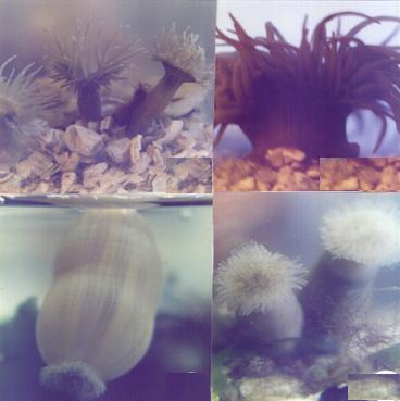
C. A Checklist of Egyptian Sea Anemones :
(Plates II, III&IV)
Family: Aiptasiidae (Carlgren, 1924).
Genus: Aiptasia
Gosse, 1858.
Species: Aiptasia diaphana (Rapp, 1829).
Family: Isophelliidae Stephenson,
1935.
Genus: Telmatactis
Gravier, 1918.
Species: Telmatactis forskalii
(Ehrenberg, 1837).
Family:
Actiniidae (Rafinesque, 1815).
Genus: Anemonia Risso, 1826.
Species: Anemonia sulcata
(Pennant, 1777).
Genus: Anthopleura Duchassaing
& Michelotti, 1860.
Species: Anthopleura stellula
(Ehrenberg, 1834).
Genus: Bunodactis verrill,
1899.
Species: Bunodactis rubripunctata
(Grube, 1840).
Species: Bunodactis verrucosa
(Pennant, 1777).
Family: Stichodactylidae Andres,
1883.
Genus: Entacmaea
Ehrenberg, 1834.
Species: Entacmaea quadricolor (Rueppell & Leuckart, 1828).
Genus: Antheopsis Carlgren, 1900.
Species: Antheopsis crispus
(Ehrenberg, 1834).
D. A Suggested Identification Key Of Sea Anemones From
1. Nematocyst-laden threads (acontia) present, sphincter muscle is
mesogleal
…………………………………………………….………..
- Nematocyst-
laden threads (acontia) absent, sphincter muscle is
endodermal
……………..…………………………..………………....
2.
Cinclides
(minute holes) usually arrange in two rows around middle
region of
body, column trumpet-shaped, common among marine
fouling of
-
Cinclides absent,
column elongated or balloon- shaped, common as a
swimming (floating) anemone as well as a usual
benthic form in
Manzalah (
……………………………………….. Telmatactis forskalii (Ehrenberg ).
3.
Body column smooth, devoid of verrucae (warts)
……..……….……..….
- Body column has outgrowths
called verrucae (warts) ……………….…...
4.
Upper edge of the column has
marginal spherules or acrorhagi (18-132
in number),
common in the intertidal region of rocky shores in
………………………………………………. Anemonia sulcata (Pennant).
-
Upper edge of the column
lacking marginal spherules or acrorhagi,
common in coral reefs of Al-Ghardaqa
(
…………………….……Entacmaea quadricolor (Rueppell & Leuckart )
5.
Verrucae are conspicuous on the upper region
of the body
column ……………………………………..………………………..……. 6
-
Verrucae
are conspicuous on the whole or most of the body
column ………..……………………………..............................................7
6.
Siphonoglyphs have red spots,
verrucae are not adhesive to any
fragments of small stones or shells debris,
common in the intertidal
region
of sandy substrates at
sea) ………………………..…………….Bunodactis
verrucosa (Pennant)
-
Siphonoglyphs lacking red spots, verrucae are adhesive to fragments of
small
stones and shells debris, common in the coral reefs at
Ghardaqa
(
…… …………….………………… ..Antheopsis crispus (Ehrenberg)
7. Marginal spherules
(4-18 in number) are present on the upper
column,
verrucae (12-24 longitudinal rows) are imperforate ,
without
red spots
and non adhesive to any fragments of
gravel, common in
fringing
reefs of AL- Ghardaqa, Sharm
el-Naga & Koseir (Red
Sea) …………………………………………………………………….
…………………….…………...…Anthopleura
stellula (Ehrenberg)
- Marginal spherules
are absent on the upper column, verrucae (24-48
longitudinal
rows) are usually perforate, with red or violet spots and
adhesive
to fragments of gravel , common in crevices on rocky shores
of
….……………………………………Bunodactis rubripunctata (Grube)
DISCUSSION
Sea anemones have been reported from the marine fouling
communities in various parts of the world seas including the English water (Orton,
1930); Coasts of America (Woods Hole Oceanographic Institution, 1952);
subtropical water of Hong Kong (Lee and Trott, 1973)
; Menai Strait, U.K (Fry, 1975) and Egyptian water (Ghobashy et al., 1980; Mona,1982 and Shoukr, 1982).
The recorded fouling anemones usually belong to the families: Diadumeniidae, Aiptasiidae, Metridiidae and Sagartiidae.
These families are acontiarians and characterized by
the presence of thread like structures called acontia
with plenty of nematocysts for offence and defence. These acontia
are shot forth through the mouth, cinclides and tentacular pores especially during contraction. These
perforations seem to act as safety valves to prevent the rupture of body during
sudden contraction, especially among fouling communities, by allowing water
jets to be squirted.
In the
The nervous system of sea anemones, in
addition to synapses with cnidae have been
studied in several species e.g. Metridium
senile, Calliactis parasitica,
Stomphia coccinea and Aiptasia pallida (Batham & Robson, 1960; Robson, 1961& 1963; Saripalli & Westfall, 1996; Westfall & Sayyar, 1997
and Westfall et al., 1998 and 1999). It is obvious that the
nervous system of Aiptasia diaphana resembles that of its allied species namely Aiptasia pallida
and Stomphia coccinea
in possessing large multipolar nerve cells in the
column. Moreover, few numbers of bipolar nerve cells have been observed among
the multipolar type. Westfall et al. (1998)
demonstrated synapses between the nerve cells and nematocysts of the sea
anemone Aiptasia pallida
and suggested a neural control of nematocyst discharge.
The swimming sea
anemone Telmatactis forskalii
was reported for the first time from Lake-Manzalah.
This anemone was unusual among sessile anemones due to its ability of free
swimming in surface water. Other anemones are known to exhibit the same
behavior such as Boloceroides hermaphroditica, Bunodeopsis medusoides and Stomphia
coccinea (Robson, 1963; Josephson
and March, 1966 and Elliott et al. 1989). However, the description of
swimming anemones from
It is worthy to
mention that the anemones are widely distributed throughout the Egyptian marine
water in
REFRENCES
Badawy, B.E. (1988): Taxonomical, histological and histochemical studies on sea anemones of the marine coasts
of
Batham, E.J. and Robson, E.A. (1960): The
nerve net of the sea anemone Metridium
senile: The mesenteries and the column. Quart. J. micr.
Sci, 101: 487-510.
Carlgren, O. (1927): Report of the Actiniaria and Ceriantharia. The
Carlgren, O. (1949): A survey of the Ptychodactiaria, Corallimorpharia
and Actiniaria. Kungl. Svenska. Veten-skapsakademiens Handlingar, (3)1(1): 1-121.
Coleman,N. (1991) :Encyclopedia of marine animals.
Dunn, D. F.(1981) : The
clownfish sea anemones : Stichodactylidae (Coelenterata, Actiniaria) and
other sea anemones symbiotic with pomacentrid fishes.
Transactions of the American Philosphical Society, 71
(1): 1-115.
EL-Ghor, A.A. (1998):
Induction of chromosomal aberrations, changes in mitotic activity and
micronuclei in mice treated with the aqueous extract of the sea anemone Parasicyonis actinostoloides.
Elliott, J.K.; Ross, D.M.; Pathirana,C. ; Miao, S.; Andersen,
R.J., Singer, p.; Kokke, W. and Ayer, W. (1989 ) :
Induction of swimming in Stomphia (Anthozoa: Actiniaria) by imbricatine, a metabolite of the asteroid Dermasterias imbricata.
Biol. Bull. 176: 73-78.
Fry,
G. W. (1975): Raft fouling in the Menai Strait,
1963-1971. Hydrobioligia,7
(3-4): 527- 558.
Ghobashy, A.F;
Ghobashy, A. F.; El-Komy,
M. and Ramadan, Sh.(1980 ) :
Fouling in the
Gohar, H.A. (1948): Commensalism
between fish and anemone. Publ. Mar. Biol. Station,
Ghardaqa,
6: 35-44.
Humason, G.L. (1972): Animal tissue techniques. 3rd ed. W.H. Francisco,
1-641.
Josephson, R.K. and March, S.C. (1966): The
swimming performance of the sea anemone Boloceroides.
J. Exp. Biol., 44:493-506.
Lee, S.W. and Trott, L .B.
(1973): Marine succession of fouling organisms in
Mona, M.H.(1982): Some
studies on fouling inhabiting some Egyptian harbors. Ph. D.
thesis, Fac. of Sci.,
Orton, J.H. (1930): Experiments in the sea on the
growth-Inhibitive and
preservative value of poisonous paints and other substances. J. Mar. Biol. Ass.
U. K., 16:373-452.
Robson, E.A. (1961): A Comparison of the nervous
system of two sea anemones, Callicatis parasitica and Metridium
senile. Quart. J. Micr. Sci., 102 (3): 319-326.
Robson, E.A. (1963): The nerve net of a swimming
anemone, Stomphia coccinea.
Quart. J.Micr. Sci., 104 (4): 535-549.
Saripalli, L.D. and Westfall, J. A. (1996):
Classification of nerve cells dissociated from tentacles of the sea anemone Calliactis parasitica.
Biol. Bull, 190: 111-124.
Schmidt,H.(1972): Prodromus
zu einer monographie der Mediterranean Aktinien.Zoologica, Wien 42 (Lieferung 2:Heft 121): 1-146.
Shoukr, F.A.M. (1982): Studies on some Coelenterates
(Cnidarians) inhabiting marine Egyptian water. Ph.D. Thesis, Fac. of Sci.,
225 pp.
Shoukr, F. A. M. (1996): A transmission electron
microscope study of cnidae and mucous cells in the
tentacles of Anemonia sulcata.
Shoukr, F. A. M. (1997): Ultrastructure
of the endosymbiont Symbiodinium
microadriaticum from a sea anemone. J. King Saud Univ., 9 (1): 25-34.
Stephenson, Y.A.
(1928): The British sea anemones. Vol. I: Ray Society,
Stephenson, T.A.(1935): The British sea anemones. Vol. II: Ray Society,
Westfall, J.A., and Sayyar, K.L. (1997): Ultrastructure of neurons and synapses in the tentacle
epidermis of the sea anemone Calliactis parasitica. J. Morph., 232:207-216.
Westfall, J.A.; Landers, D.D. and McCallum, J.D.
(1998): Different nematocytes have different synapses
in the sea anemone Aiptasia pallida (Cnidaria, Anthozoa). J. Morph., 238: 53-62.
Westfall, J.A., Landers, D.D. and McCallum, J.D.
(1999): Ultrastracture of neuro-spirocyte
synapses in the sea anemone Aiptasia pallida (Cnidaria, Anthozoa, Zoantharia). J. Morph., 241: 165-173.
Woods Hole Oceanographic Institution (1952): Marine
fouling and its prevention.
Explanation of Figures and
Plates
Fig. 1 : T.S. of a tentacle of Aiptasia diaphana
showing perforation and
tentacular retractor muscle.
Fig. 2 : L.S. through a soft spot showing
incipient cinclide.
Fig. 3 : T.S. of the column showing the
general histological structure.
Fig. 4 : L.S. of the upper part of the column
showing ectodermal longitudinal
muscles
and mesogleal sphincter.
Fig. 5 : L.S. of the pedal disc showing the
basilar muscle.
Fig. 6 : T.S. of a female mesentery showing a
circumscript-diffuse retractor
muscle.
Fig. 7 : T.S. of a mesenteric filament
showing the general histological structure.
Fig.8 : T.S. of an acontium
showing large-sized nematocysts.
Fig.9 : Arrangement of mesenteries.
(A): Octamerous, with 8 perfect mesenteries
including 3 directives.
(B): Heptamerous, with 7 perfect mesenteries
including 3 directives.
(C): Hexamerous, with 6 perfect
mesenteries including 2 directives. (Black rectangles = directive
mesenteries)
Fig. l0: Nematocyst signature.
Tentacles
(A, B, C); column (D, E); actinopharynx (F, G);
mesenteric
filaments (H, I) and
acontia (j).
Plate I
Fig. A: T.S. of the column ectoderm of Aiptasia
diaphana showing the ectodermal
nerve
net
(silver impregnation, x 1000).
Fig. B: T.S. of the column endoderm showing the endodermal nerve net (silver
impregnation, x 1000).
Fig.
C: T.S. of a mature ovum in a female anemone showing absence of trophonema
(Haematoxylin-eosin,
x 1000).
Fig. D: T.S. of a sperm follicle in a male anemone
showing spermatogenesis
(Haematoxylin-eosin, x 400).
a.t., alveolar tract; ac., acontium;
agr.c., agranular cell; b.m., basilar muscle; bp.n.c.,
bipolar nerve cell; c.m.f., circular muscle fibers; ci.t., ciliated tract; cn., cinclide; cn.g.t., cnidoglandular tract, co.ep.c., columnar epithelial cells; ec.,
ectoderm; en., endoderm; ep.c., epithelial cells; g.c, gland cells ; gr.c.,
granular cells; in.c., interstitial cell; l.m.f., longitudinal muscle fibers;
m., mesoglea; m.f. mesogleal fibers; m.sp., mesogleal sphincter; me.,
mesentery; me.f., mesenterial
filaments; mp.n.c., multipolar
nerve cell; n.nt., nerve net; nc.,
nematocysts; nl., nucleolus; nu.,
nucleus; ov., ovum; p.m., parietobasilar
muscle; r.m., retractor muscle; s.c.,
sensory cell; sc., spirocysts; spc.,
spermatocyte; spg. spermatogonia; spz.,
spermatozoa; t.r.m., tentacular
retractor muscle; y.g., yolk granules; z., zooxanthella; z.t. zooxanthella tract.
Plate II
Fig.1
: Aiptasia diaphana showing acontia
&cinclides.
Fig.2
: Telmatactis forskalii showing :
A:
swimming form, B&C: Benthic forms.
Plate III
Fig.1 : Anemonia
sulcata. Fig.4:
Bunodactis rubripuctata.
Fig.2 : Anthopleura
stellula. Fig.5:
Bunodactis verrucosa.
Fig.3: Entacmaea quadricolor. Fig.6: Antheopsis crispus.
Plate IV
Fig. A: The
benthic forms of Telmatactis forskalii.
Fig. B: The
swimming form of Telmatactis forskalii.
Fig. C: The
green tentacled form of Anemonia
sulcata.
Fig. D: The
brown tentacled form of Anemonia
sulcata.
a., acontia ; ac., acrorhagi ; c., cinclides ; co., column ; 1.s. longitudinal striations ; o.
d., oral disc ; p. d., pedal disc (base) ; r., red spot ; s., siphonoglyph ; st., stomodaeum; s.f., symbiotic fish,
t., tentacles ; v., verrucae.
Table (1): Relation between numbers of tentacles, cinclides,
mesenteries at base and disk with the anemone size
(base diameter) in eight specimens of Aiptasia
diaphana.
|
base
diameter (mm) |
No. Tentacles |
No. Cinclides |
No. of
mesenteries (pairs) |
|
|
at base |
at disk |
|||
|
2 4 6 8 10 12 18 20 |
29 75 95 100 98 95 114 114 |
7 16 22 26 32 35 35 38 |
40 42 42 43 46 45 46 46 |
28 38 40 41 42 40 42 43 |
Table (2): Histochemical
tests affinities to cnidae and
zooxanthellae of Aiptasia diaphana.
|
Histochemical tests |
Cnidae |
Zooxanthellae |
|
|
Spirocysts |
Nematocysts |
||
|
PAS Alcian blue (pH.2.5) Mercurice bromophenol blue |
+ + + + + |
+ + - + + |
+ + + + + + + + |
+ + + strong reaction
+ + Moderate reaction
+ Weak reaction
- negative
reaction
Table (3): Comparison
between the sea anemone species identified from the Egyptian marine water.
|
Anemone
species |
Locality
of specimens |
Base
diameter range (mm) |
Column
length range (mm) |
Number
of tentacles range |
Number
of mesenteries range |
|
Aiptasia diaphana |
Port-Said,
Lake-Timsah, |
2-20 |
5-40 |
29-114 |
28-46 (pairs) |
|
Telmatactis forskalii |
Lake-Manzalah ( |
3-15 |
20-120 |
24-48 |
24-48 (pairs) |
|
Anemonia
sulcata |
|
1-41 |
2-26 |
24-250 |
28-80 (unpaired) |
|
Anthopleura stellula |
Al-Ghardaqa, Koseir Sharmed-Naga ( |
3-20 |
5-30 |
22-48 |
18-30 (pairs) |
|
Banodactis rubripunctata |
Alexandria
in Abou kir, Rasel-Tin & El-Dekhela |
2-50 |
4-30 |
24-96 |
24-48 (pairs) |
|
Banodactis verrucosa |
|
3-35 |
4-27 |
84-96 |
24-48 (pairs) |
|
Entacmaea quadricolor |
Al-Ghardaqa ( |
18-100 |
12-115 |
76-300 |
50-85 (pairs) |
|
Antheopsis crispus |
Al-Ghardaqa ( |
25-70 |
20-130 |
88-285 |
62-90 (pairs) |
Prof. Dr. Fayez Shoukr, Professor of Invertebrates, Zoology
Department, Faculty of Science, Tanta University, Tanta 31527,Egypt.
Shoukr, F.A., Mona,
M.H. and Badawy, B.E. (2000) : Taxonomy Of Some Sea
Anemones (Cnidaria: Anthozoa)
From
تصنيف بعض شقائق البحر ( اللاسعات :
الزهريات )
في مصر
د. فايز عبد المقصود شكر ،د. محمد حسن منا ،د. بدوي السيد
بدوي
قسم علم الحيوان ، كلية العلوم ، جامعة طنطا ، مصر
أظهرت هذه الدراسة انتشار شقائق البحر
انتشاراً واسعاً على جوانب السفن الراسية في قناة السويس وخاصة في ميناء بورسعيد
وبحيرة التمساح كأحد مكونات الحشف البحري. وقد أستدعى ذلك دراسة
الوصف التصنيفي لأحد الأنواع الشائعة وهو ابتازيا ديافانا لأول مرة في مصر . وقد تبين
من الدراسة التشريحية وجود الخيوط اللاسعة التي تنطلق من خلال فتحات دقيقة في جدار
الجسم، وأن عدد المسا ريقا يتراوح بين 28-46 زوج تنوع ترتيبيها من النظام السداسي
الشائع إلى السباعي أو الثماني نظراً لشيوع التكاثر اللاجنسى. وأوضحت الدراسة الهستولوجية أن العضلات العاصرة ضعيفة جداً ومندمجة في الميزوجليا وعضلات المسا ريقا تتميز إلى عضلات مرجعة طولية من
النوع الممتد البسيط وأخرى جانبية قاعدية ضعيفة والعضلات القاعدية بسيطة. وتوجد
شبكة عصبية على الجزء الداخلي من طبقة الاكتودرم تتكون
من خلايا عصبية ثنائية وعديدة الأقطاب وألياف عصبية، وكذلك شبكة عصبية أندودرمية تشبه نظيرتها في طبقة الاكتودرم.
و أوضحت الدراسة الهستوكيميائية أن الخلايا اللاسعة
تحتوى على المواد البروتينية المرتبطة بالمواد عديدة السكاكر وهى المكون الأساسي للمادة اللاسعة داخلها.
وبالإضافة إلى ذلك فقد تم تسجيل أحد
شقائق البحر السابحة لأول مرة في بحيرة المنزلة بالقرب من بورسعيد، وكذلك عرض
قائمة تصنيفية بالأنواع التي تم وصفها حتى الآن واقتراح مفتاح تصنيفي للتفرقة بين
الأنواع المختلفة من شقائق البحر وتوضيح أماكن معيشتها.